Categories


- 0086-512-58697986
- [email protected]
- bestrantech
- 0086-13862268429
High Frequency Digital Radiography System

BT-XR22 High Frequency Digital Radiography System
I. Application
BT-XR22 Digital high frequency medical surgical X-Ray equipment can meet different parts radiography, such as head, chest, abdomen, waist, lumber, thoracic, pelvis, limbs etc. And meet different positions radiography, such as lay decubitus, normotopia, side position etc.
II. Specification
|
Item |
Content |
Technical Parameters |
|
|
Power supply |
Voltage |
380V±38V |
|
|
Frequency |
50Hz±1Hz |
||
|
Capacity |
≥63kVA |
||
|
Internal Resistance |
≤0.17Ω |
||
|
High Voltage Generator (SEDECAL) |
Power |
50KW |
|
|
Inverter Frequency |
60 KHz |
||
|
Tube voltage |
40kV—150kV |
||
|
Tube current |
10mA—640mA |
||
|
mAs |
0.1mAs—10000mAs |
||
|
X-ray tube (TOSHIBA) |
Tube Focus: |
0.6 /1.2mm |
|
|
Output Power |
Big focus: 50kW Small focus: 20kW |
||
|
Anode thermal capacity |
210KJ |
||
|
Digital Work Station |
Computer System |
Brand: DELL OPTIPLEX 7010 commercial use Processor: Core™ i5-3570 processor RAM: 4GB DDR3 Hardware: 500GB SATA (7200 rpm) CD-driver: suit MT’s 16X variable-speed DVD + / - RW With functions of double written Network Card: Broadcom NetXtreme 10/100/1000 PCIe Gigabit LAN GNTB-A High display card: 1GB AMD RADEON HD 7570,FH,with DVI-VGA Adapter Slop: PCIE seriel port and parallel port, all height, MT |
|
|
Color LCD Monitor |
19 inches 1M LED backing light LCD display revolution1280x1024, 5:4screen dot pitch: 0.294mm contrast ratio: 1000:1 brightness: 250cd/㎡ Grey feedback time: 5ms visual Angle: 160/170° |
||
|
1. Image playback: thumbnails view, sequence replay tools. 2. Image processing: W/L adjustment, arrows and words etc labels, angle and distance measurement, image scaling, translation, up/down conversion, left/right conversion, rotation, black and white reversal. 3. Image storage: image real-time storage, DICOM image send, A shampoo Burning Studio, derived storage (choose various storage ways Bitmap, JPEG, AVI etc to be used in Word and Power-point office software, it is convenient for the doctors to write diagnostic reports and papers ). 4. DICOM3.0: can be connected with laser camera to print film and PACS NETWORK. 5. Medical record management: database management, graphic report, support for WORKLIST. |
|||
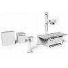
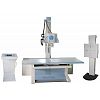
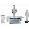
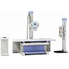
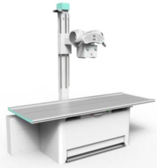
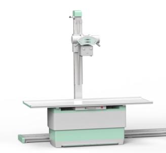
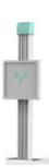
III. Table and Bucky stand
|
Item |
SYC20 |
SYC30 |
XPJ30 Bucky stand |
|
Picture |
|
|
|
|
SID |
500-1280mm |
500-1280mm |
|
|
X-ray tube Rotation |
±90° |
±90° |
|
|
X-ray tube Longitudinal movement |
≥1300mm |
≥2500mm |
|
|
Pillar Rotation range around |
±180° |
±180° |
|
|
Table transverse movement |
≥220mm |
≥220mm |
|
|
Table longitudinal movement |
≥900mm |
≥900mm |
|
|
Cassette Longitudinal movement |
500mm |
500mm |
130mm |
|
Height |
720mm |
500mm~720mm |
450mm~1780mm |
|
Grids(JPI) |
Grid density: 103L/inch Grid ratio: 10:1 Focusing Distance: 100cm Stationary type: 15"×18" |
Grid density: 103L/inch Grid ratio: 10:1 Focusing Distance: 180cm Stationary type: 15"×18" |
|
IV. Flat panel detector
|
|
Toshiba FPD |
Toshiba Portable FPD |
CareView FPD |
|
Active area |
430(H)×439(V) |
350(H)×430(V) |
430(H)×430(V) |
|
Pixel matrix |
3008(H)×3072(V) |
2448(H)×2984(V) |
2816(H)×2816(V) |
|
Pixel pitch: |
143 μm |
143 μm |
154 μm |
|
Cycle time: |
Less than 6 s |
6s |
9s |
|
Limiting resolution |
3.5 lp/mm |
Min. 3.7 lp/㎜ |
Min. 3.5 lp/㎜ |
|
A / D transition: |
14 bit |
16 bit |
14 bit |
|
Energy range: |
40 - 150 kVp |
40 - 150 kVp |
|
|
Max entrance dose: |
4 mR / frame |
4 mR / frame |
|
|
Data output: |
16 bit |
16 bit |
16 bit |
|
External material: |
Carbon Fiber |
Carbon Fiber |
Carbon Fiber |
V. Features
1) SEDECAL high voltage generator and TOSHIBA X-ray tube provide constant direct current and high voltage output, will achieve high quality monochromatic X-Ray and eliminate the harmful effect of soft ray.
2) Equipped with high performance and large capacity X-Ray tube, adopt 0.6/1.2mm dual focus, 300KHU large capacity and high speed X-Ray tube, it is suitable for long time and high intensity clinical examination.
3) Imported /domestic DR flat panel detector connected with image processor achieves low noise and rich contrast image, edge enhancement filtering device makes the edge of image more clear and sharp.
4) Digital workstation, fast image processing, connected DICOM3.0.
5) Intelligent high voltage control system with multiple human APR, LCD display show APR rules, and set according patients figure, position and part automatically, it’s fast and convenient.
6) Computer progress control, remote operate, electric multi-lead collimator, it’s easy to adjust X-ray range, this reduce X-ray harm for patients and atmosphere.
7) With IBS function and four choices to set KV and mA diagram. AEC offer accurate exposure.
8) Multi self-diagnosis program, has RS232 port, and with error indication function.
9) Comfortable, convenient and exquisite table, two beds for options, SYC30 can give long range, this promise wheelchair and stretcher radiography and more up and down range, this way is more convenient for small figure.