Categories


- 0086-512-58697986
- [email protected]
- bestrantech
- 0086-13862268429
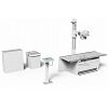
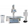
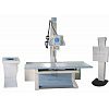
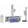
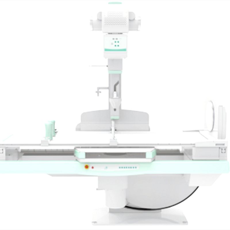
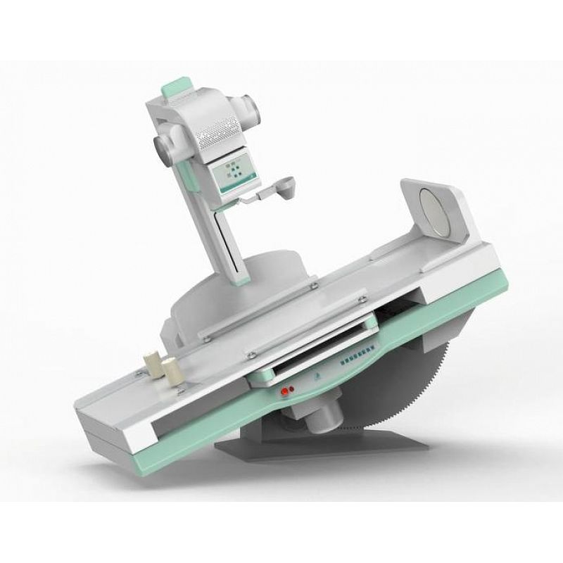
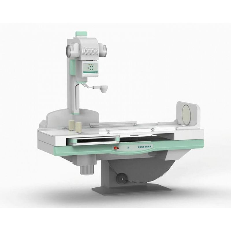
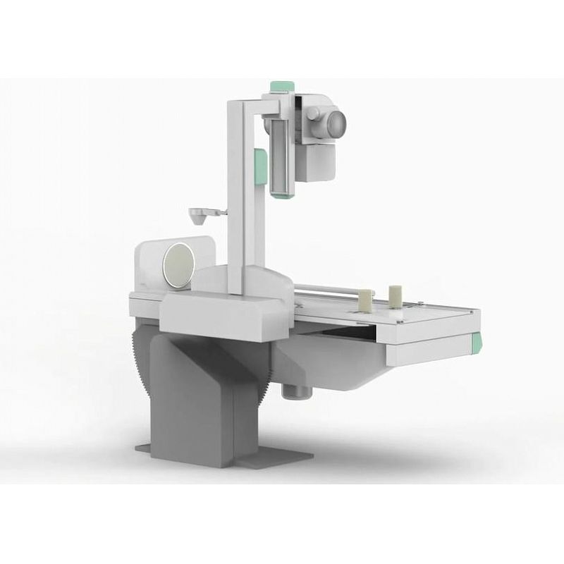
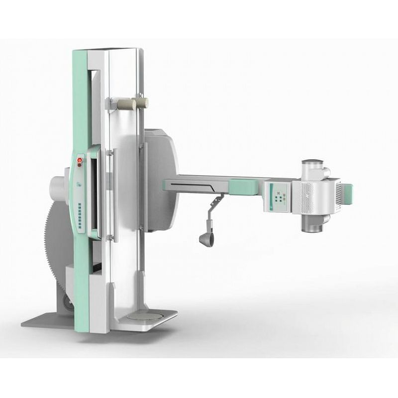
BT-XF19B Medical Diagnostic X-ray System
II. Standard Configuration:
1 | Console | KZT40 | 1Unti |
2 | Generator | FSQ80 | 1Unit |
3 | X-ray tube | E7869X | 1Unit |
4 | Collimator | XSQ20 | 1Unit |
5 | Diagnostic table | ZDC20 | 1Unit |
6 | Image intensifier | E5830SD-P6A | 1Unit |
7 | CCD Camera | acA1920 | 1Unit |
8 | Digital image process system | DRFOC | 1Unit |
9 | Flat Panel Detector | PLD1417V | 1Unit |
10 | Medical use display | U2412Mc | 1Unit |
11 | Cables and accessories | PLD8600 | 1Unit |
III. Specifications:
Item | Content | Technical Parameters |
Power Supply | Voltage | 380V±38V |
Frequency | 50/60Hz±1Hz | |
Capacity | ≥105kVA | |
Internal Resistance | ≤0.11Ω | |
High frequency high voltage x-ray generator | Power | 80kW |
Inverter Frequency | 440 kHz | |
Tube voltage | 40kV—150kV | |
Tube current | 10mA—100mA | |
mAs | 1.0ms—10000ms | |
Time | 0.1 mAs~1000 mAs | |
Fluoroscopy Tube Voltage | 40kV—125kV | |
Fluoroscopy mA | 0.5mA—10mA/ 5mA- 20mA | |
X-ray tube assembly | Tube Focus: big/small | 1.2mm /0.6mm |
Input power | Big Focus/100kW Small focus/40kW | |
Anode thermal capacity | 600KHU | |
Anode Rotation Speed | 9700rpm | |
Micro-computer control digital tube remote diagnostic table
| Table rotate Movement range | 90°~0°~-25° |
Table transverse movement distance | ±110mm | |
Longitudinal movement distance of radiography frame and spot film device | ≥720mm | |
SID | 1100- 1500 mm | |
X-Ray tube focus to film distance | 8″×10 ″- 14″×17″ | |
Collimator | Full film, half dividing, three parts dividing, four parts dividing | |
Fixed grid for table use | 80N ~ 130N | |
Table rotate Movement range | ±360º Infinite rotation | |
Table transverse movement distance | Electric multi-leaf | |
Longitudinal movement distance of radiography frame and spot film device | Grid density: 103L/INCH, Ratio: 10:1, Focusing distance: 130cm, stationary type :15” ×18” | |
Flat Panel Detector | Toshiba fixed Flat Panel Detector | Limiting resolution: 3.4 lp/mm typ Vision size: 430(H)×439(V) Pixel Matrix: 3008(H)×3072(V) Pixel gap: 143 μm A / D transform: 14 bit |
DR Software | DRFOC | Basic operation: change control console password, edit ID, acquisite images. Additional operation: Add new check, edit present checking info, add new position, change image aquisition order, multi-checking agreement chance, manual adjustment of exposure parameters, automatic exposure control mode, focus choice, patient body-type choice, tube capacity check, ESA curve choice, image cutting, note added (sent to DICOM workstation), mark on images, rotate or overturn, full-size image observation, check patient info and dose info, accept or refuse images. Image management: change order, patient basic info editing, inquiry history images, resend history images, re-print history images, check images mark info, review history images, manage refused images, space reclaimed, image protection, and manual image deletion etc. System management: ID edition, change ID password, ED refrigeration set, statistics info checking, detector calibration, equipment control, output order management, image measurement. |
Digital Image TV System | Image Intensifier | 230mm |
high resolution CCD | ultra-low-light, standard 2 mega-pixel | |
Digital Picture Processing System (CCU Sentinel) | High definition line by line output mode, 8 level noise reduction, store 8 image; LIH (freeze the last frame), the image can be turned vertically and horizontally, positive and negative image; OSD(monitor show) | |
Gray scale display 19’’ | resolution ratio:1280x1024 Luminance: 1000cd/㎡ |
IV. Features
l The floating table with bucky stand can meet the photographic requirements of different standing and lying positions. The bed floating and electromagnetic lock design makes it convenient for the accurate position of the lying patient, operation is much more convenient and flexible.
l Digital flat Panel detector can help you get the high-definition images.
l The leading domestic high power compact high frequency X-ray generator and high frequency power inverter makes the machine much more compact and more convenient without the extra high-voltage generator and cable.
l The application of the KV and MA digital closed-loop control technology and the real time control of the microprocessor ensure the accuracy and repeatability of the dose.
l The multiple automatic protection features and fault tips ensure safety during the operation process.
l The system has excellent performance and high quality images, from the fluroscopy,spot film, sequence of all digital photography collected to digital subtraction angiography (DSA) image processing.
l The table can be +90 degrees to 0 to -25 degrees rotate.
l Humanized design of diagnosis bedside switch operation, can control the table body and imaging system movement,so that the close table inspection is convenient and easy operation.
l Spot film device and imaging system movement range more than 720 mm.
l Adopts the operation of the machine move, but the patient don't need move, can easily finish from the throat, esophagus to the lower abdomen of a series of inspections.
l Cassette trolley can test cassette size by itself, a key can completely finish the insert piece, save space, convenient and fast.