Categories


- 0086-512-58697986
- [email protected]
- bestrantech
- 0086-13862268429
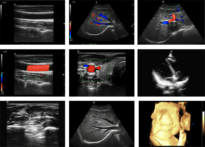
4D Portable color doppler machine
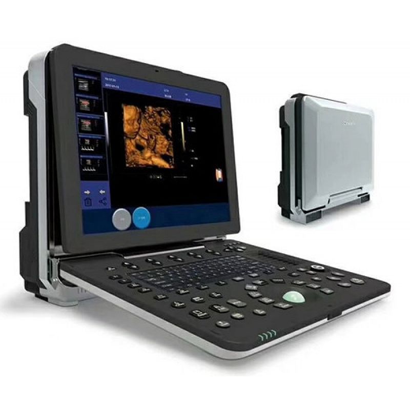
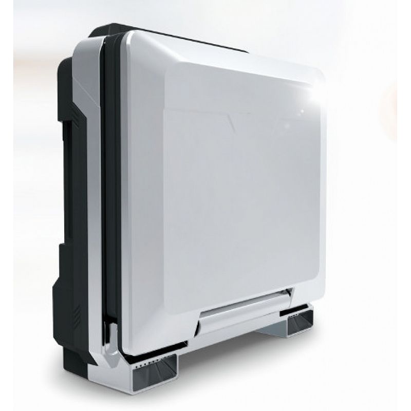
4D Portable color doppler machine
|
1 |
A summary of the main specifications and system of portable color Doppler ultrasound |
|
1.1 |
Portable all digital color Doppler ultrasonic host |
|
1.2 |
Ultrasonic host operating system: Windows 8 operating system |
|
1.3 |
Spectrum pulse Doppler |
|
1.4 |
Direction energy Doppler |
|
1.5 |
Real time three synchronization |
|
1.6 |
Space composite imaging: the requirement is 3 level, visual adjustable. |
|
1.7 |
Organized harmonic imaging technology |
|
1.8 |
4B imaging mode |
|
1.9 |
One key intelligent optimization |
|
1.10 |
Chinese and English interface, Chinese and English input, optional |
|
1.11 |
Monitor: 15 inches, high definition ultrasonic display liquid crystal display |
|
1.12 |
Display 0-30 degree angle adjustable |
|
1.13 |
Physical clipboard: save the image on the left side of the screen, which can be directly saved or deleted. |
|
1.14 |
The system has the function of on-the-spot upgrade |
|
1.15 |
Presupposition: for different inspection of the viscera, preset the inspection conditions for the best image, reduce the adjustment of the operation, and the commonly used external adjustment and combination regulation. |
|
1.16 |
Support real-time 3D imaging function |
|
1.17 |
The probe interface is 2 |
|
2 |
probe |
|
2.1 |
Convex array probe: fundamental frequency 2.5MHz/3.0MHz/3.5MHz/4.0MHz/H4.0MHz/H5.0MHz, six Duan Bianpin |
|
2.2 |
Linear array probe: fundamental frequency: 6.0MHz/7.5MHz/8.5MHz/10.0MHz/H10.0MHz, five stage frequency conversion. |
|
2.3 |
Transvaginal probe: fundamental frequency: 4.5MHz/6.0MHz/7.0MHz/9.0MHz/H8.0MHz, five stage frequency conversion. |
|
2.4 |
Phased array probe: 2.5MHz/3.0MHz/3.5MHz/4.0MHz/H3.0MHz/H4.0MHz, six stage frequency conversion |
|
2.5 |
Micro convex probe: 4.5MHz/6.0MHz/7.0MHz/9.0MHz/H8.0MHz five stage frequency conversion |
|
2.6 |
Volume probe: 2.0MHz/3.0MHz/4.5MHz/6.0MHz/H5.0MHz, five stage frequency conversion |
|
2.7 |
The above probe has harmonic frequency |
|
2.8 |
It can be selected according to customer needs: convex array probe, linear array probe, transrectal probe, micro convex probe, phased array probe, volume probe. |
|
2.9 |
Under each probe, there is a selection of specialist and viscera mode and rapid entry detection. |
|
3 |
Two-dimensional imaging mode |
|
3.1 |
Gain: 0-100, step 1 visible adjustable |
|
3.2 |
TGC: 8 segment adjustable |
|
3.3 |
Image optimization: visible and adjustable over 7 levels |
|
3.4 |
Dynamic range: 20-280dB 20 level visual adjustable (maximum value and adjacent step image proofs). |
|
3.5 |
False color: 12, visible and adjustable |
|
3.6 |
Smooth treatment: 8, visible and adjustable |
|
3.7 |
Edge enhancement: 8, visible and adjustable |
|
3.8 |
Sound power: 5% to 100%, step 5%, visible and adjustable |
|
3.9 |
Display depth: greater than 317mm, less than 20mm, max depth of probe 371mm |
|
3.10 |
Maximum focus number: 6 focal points, which can be moved throughout the whole process. |
|
3.11 |
Scan line density 256 visible tunable |
|
3.12 |
Gray scale: 0-7 level visible visible and adjustable |
|
3.13 |
Filtering, 5 kinds |
|
3.14 |
Scanning range, 50%-100% |
|
3.15 |
Frame correlation, 0-4 level, visible and adjustable |
|
3.16 |
The screen has 14 forms of real time display of voice power, probe frequency, dynamic range, pseudo color, grayscale and so on. |
|
4 |
Color imaging mode |
|
4.1 |
Color frequency: 8 frequency conversion, visible adjustable |
|
4.2 |
Color deflection: equipped with |
|
4.3 |
Color frame correlation 12 levels, visible and adjustable |
|
4.4 |
Color map: 7, visible and adjustable |
|
4.5 |
Color reversal: adjustable |
|
4.6 |
B/C split screen synchronous display function: equipped with |
|
4.7 |
Color baseline: 7, visible and adjustable |
|
4.8 |
Color line density: adjustable |
|
5 |
Spectrum Doppler mode |
|
5.1 |
Sampling volume angle correction: -80 degree to 80 degree adjustable |
|
5.2 |
Sampling volume: 0.5mm-20mm visibility adjustable |
|
5.3 |
Frequency: ≥5, visible and adjustable |
|
5.4 |
Baseline: 8 adjustable |
|
5.5 |
Smooth: 8 files can be adjusted |
|
5.6 |
Display layout: 4 visible tunable |
|
5.7 |
False color: 7 kinds of adjustable |
|
5.8 |
Speed scale: 3-2288cm/s |
|
5.9 |
Spectrum envelope function: real time automatic spectrum envelope, manual spectrum envelope, and so on. System automatic analysis and display: PSV, EDV, RI, PI, S/D, ACC, HR and other kinds of data |
|
6 |
Measurement and analysis function: |
|
6.1 |
General measurement distance, area, angle, time, slope, heart rate, velocity, acceleration, neck hyaline layer, spectrum tracing, resistance index / pulsatility index, etc. |
|
6.2 |
Obstetric measurement: weight measurement formula 8 options |
|
6.3 |
The color and line type of the measuring line can be adjusted at will (including activating the color and completing the color). |
|
6.4 |
The measurement results show that the location and font size can be adjusted according to needs. |
|
6.5 |
Professional software package: abdomen, volume, ratio, obstetrics and Gynecology, small organs, carotid, Urology, orthopedics, peripheral blood vessels, heart. |
|
7 |
Graphic and text management system |
|
7.1 |
Host built in ≥128G solid state hard disk to start fast and stable |
|
7.2 |
Movie playback: 1200 frames |
|
7.3 |
Internal patient file information management system: can record patient number, name, check number, check date and so on, and can be searched and managed by numbering, checking number, name and so on |
|
7.4 |
The type of report is 16 |
|
7.5 |
One key fast report graphic and text management |
|
8 |
Interface |
|
8.1 |
USB interface: 2 |
|
8.2 |
VGA interface: 1 |
|
8.3 |
COME interface: 1 |
|
8.4 |
LAN interface: 1 |
|
8.5 |
HDMI interface: 1 |
|
9 |
To configure |
|
9.1 |
Portable full digital color Doppler ultrasound diagnostic system host 1 sets |
|
9.2 |
Probe: convex array probe (marking), high frequency probe (selection), overcast probe (selection), heart probe (selection), volume probe (selection), medical trolley (selection) |
|
10 |
Technology, after-sales service and other requirements |
|
10.1 |
After acceptance, the warranty is free for two years |
|
10.2 |
Manufacturer has ISO13485 certification and EU CE certification. |